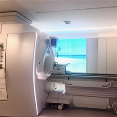
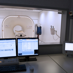
Hybrid PET/MR imaging is an advanced technique that simultaneously combines two complementary modalities: Positron Emission Tomography (PET) and Magnetic Resonance Imaging (MRI). This integration enables the acquisition of both metabolic and molecular data from PET and high-resolution anatomical and functional information from high-field MRI in a single study.
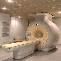
This technology represents a major advancement in medical imaging, at both the clinical and preclinical levels, providing greater accuracy in localizing regions of abnormal metabolic activity, superior tissue contrast, and advanced functional assessment for the simultaneous study of metabolism, perfusion, diffusion, and structure.
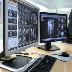
Hybrid PET/MR imaging is a key tool for translational biomedical research, advanced diagnostics, and the evaluation of innovative therapies.