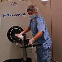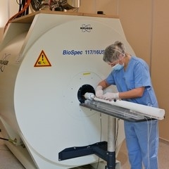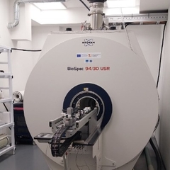
PET/MRI 9.4T Bruker BioSpec with CryoProbe
High-field Magnetic Resonance Imaging (MRI) system operating at 9.4 tesla, enabling the acquisition of both anatomical and functional images with exceptional resolution. The system supports in vivo MR spectroscopy, angiography, and cardiovascular, perfusion, and diffusion studies.
Its modular design, featuring gradient systems and RF coils of various sizes, makes it highly optimized for a wide range of preclinical MRI applications.
The system includes a CryoProbe, providing a sensitivity gain of 2.5 to 5.3 times compared with standard room-temperature RF coils. This enhanced sensitivity delivers key anatomical and functional information for in vivo preclinical studies.
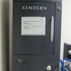
It also includes a PET insert, as described in the section on Hybrid MRI/PET Imaging, enabling the simultaneous acquisition of metabolic and structural data.
This high-field MRI system has been acquired by BioImaC with funding from the European Union through the NextGenerationEU program.
Location: BioImaC